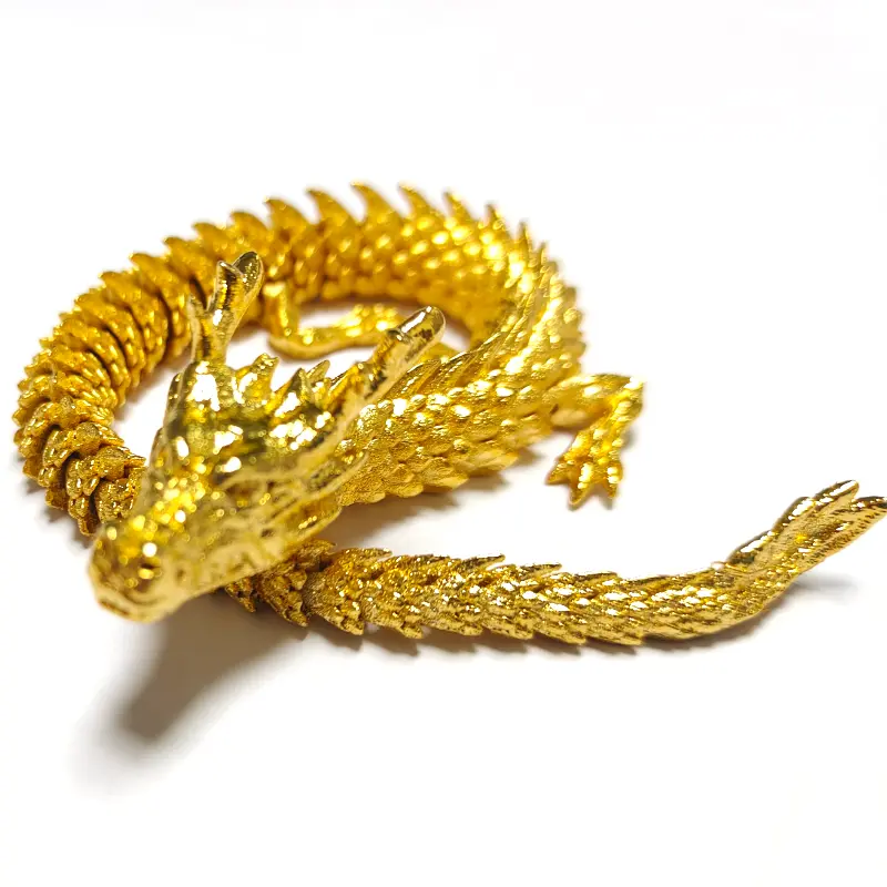
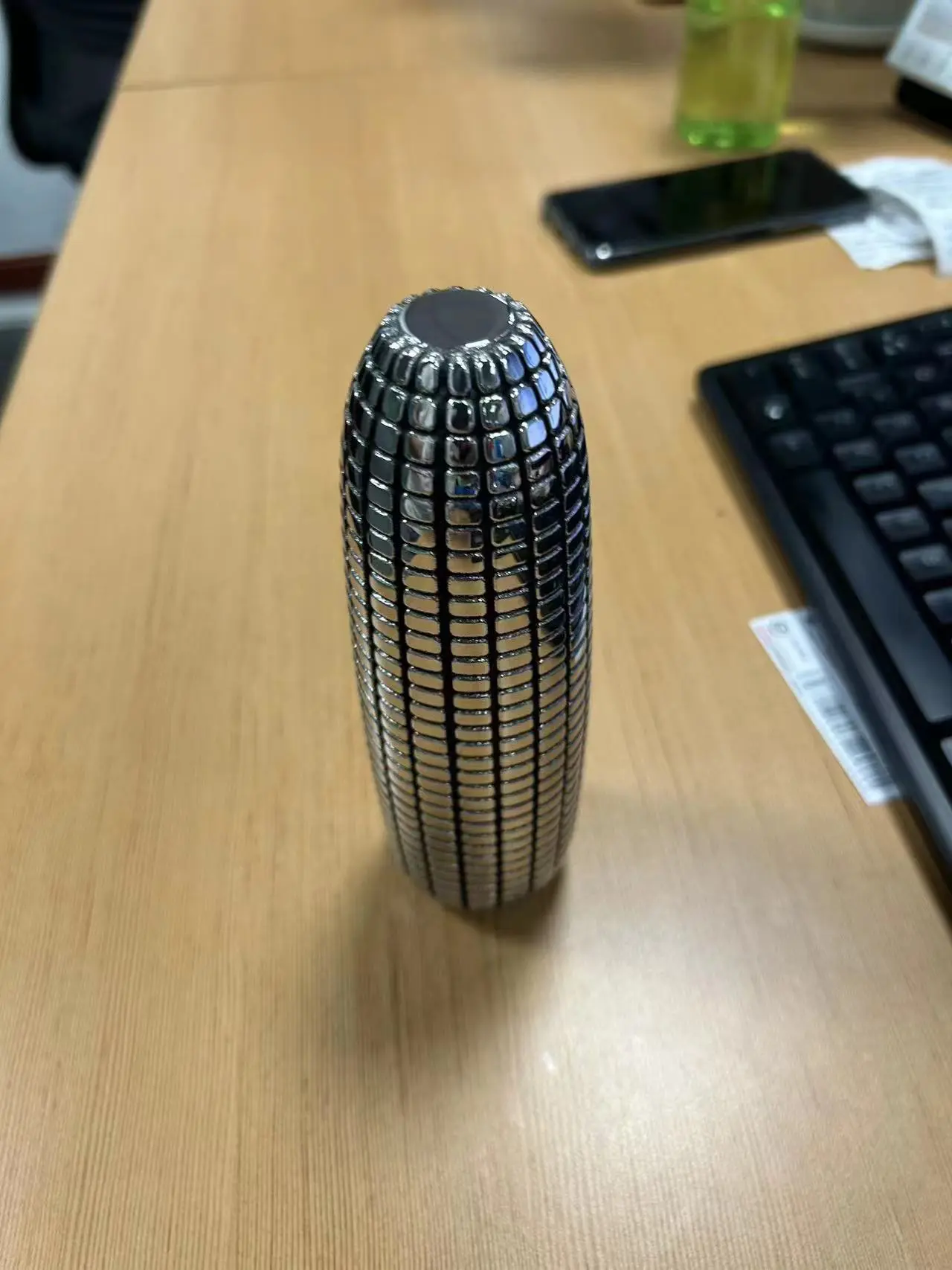
Recently, according to the Resource Library, the Research Team of the Eindhoven University of Technology (Tu / E) has successfully applied a new 3D printing technology based on optics – Xolography – to the printing of live cells. This breakthrough lays the basics of the printing of complex organic tissue such as kidneys and muscles. The researchers have succeeded in printed structures which are roughly the size of human cells and are as small as 20 microns.
Related search results are published in the journal Advanced Materials. Although technology is always at the experimental stage, researchers think that this is an important step in the field of tissue engineering in the future.
Innovation in Xolography technology
The Tu / E team has developed a compact prototype prototype based on Xolography technology, which is equivalent to a suitcase. This technique allows printing by exposing the liquid polymer to a transverse beam.
Lena Stoecker, a doctoral student from the biomaterial and bio-fabrication group, directed the study, working to optimize the method of biomedical applications. His work focuses on the development of materials and technologies that can strongly simulate the natural cellular environment, a major challenge in tissue engineering.
Unlike traditional 3D layer printing, Xolography simultaneously solidifies the entire volume by projecting a series of images in the photoreactive fluid. This approach allows the printing of faster and higher resolution, but requires significant improvements to technology to support living cells. The research team has developed biocompatible materials that have replaced toxic photoinitiators to ensure the safety of biomedical applications.
Research results and application potential
Stoecker has successfully printed hydrogel scaffolding with pores of 100 micron at 1 mm which allow nutrients to sink to support cell culture. In addition, the researchers obtained precise control of the properties of the materials by adjusting the light intensity, allowing the creation of structures with a different hardness.
A major breakthrough is the use of thermally sensitive hydrogels to reach the shape of the structure over time (that is to say 4D printing). This innovation opens the way to the manufacture of artificial muscles which can contract and develop with temperature changes.
Although the clinical application takes some time, the team’s research has thrown an important basis for the future of biopritation. Other developments can lead to more complex tissue models for drug tests, thereby reducing dependence on animal experiences.
In addition, the ability to print highly personalized implants and regenerated fabric structures can revolutionize personalized medicine, providing new solutions for organ failure and trauma recovery.


